Abstract
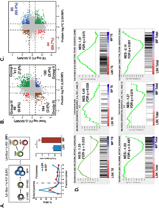
Mechanisms of translational regulation are poorly understood in hematopoietic stem cells (HSCs) and committed progenitors (MP). In order to investigate the impact of translational regulation on early mouse hematopoietic development, we characterized the translatome using a combination of polysome profiling, RNA-sequencing (RNA-seq) of polysome-associated and total cellular mRNA, and whole proteome evaluation. Comparison of RNA-seq data from HSC-enriched LSK (Lin-Sca-1+c-Kit+) and MP (Lin-Sca-1-c-Kit+) cells showed more differentially expressed mRNAs in polysomal RNA than total RNA (412 vs 280 mRNAs, respectively) among ~15,000 mRNAs analyzed. In addition, polysomal mRNAs were enriched for a unique set of functional pathways (e.g. inflammatory response, apoptosis and p53) compared to total RNA in both LSK and MP cells. Interestingly, although LSK cells showed ~20% lower global translation than MPs as demonstrated by polysome profiling (Figure 1A), they exhibited significantly higher translational efficiency (TE = polysome RNA abundance/total RNA abundance) for mRNAs that support HSC maintenance (e.g. glycolysis, fatty acid metabolism, oxidative phosphorylation, mTOR signalling). Integration of proteomic and RNA-seq data demonstrated that 605 out of 784 (77.2%) differentially expressed genes (DEGs) between LSK and MP cells identified based on total RNA-seq data (Groups I & III; Fig 1B) also showed a corresponding change in protein expression, while remaining 179 DEGs (22.8%; Groups II & IV; Fig. 1B) showed an anti-correlation. Remarkably, in the latter group, expression of 129 proteins (72.1% of all differentially expressed proteins in LSKs vs MPs) correlated with their TEs (Figure 1C). While gene set enrichment analysis of published HSC regulators showed an enrichment in LSKs in total RNA-seq data, such an enrichment was not observed when evaluating mRNAs with differential TEs. However, mRNAs with high TE confirmed a surprising enrichment in mTOR-responsive genes independent of their total RNA expression in LSKs, but not in MP cells (Figure 1D). To investigate the biochemical basis of this observation, we performed western blot analysis of LSK and MP cells and observed decreased mTOR protein expression and signaling in purified MP cells, despite their higher global levels of translation. In addition, despite abundant expression of mTOR protein in LSK cells, 4E-BP1, a known target of mTOR, was only phosphorylated at the priming residues Thr-37/46 but not at the downstream Ser-65, a residue that initiates cap-dependent translation. In contrast, MP cells phosphorylated Ser-65, consistent with its increased translation despite the absence of mTOR signalling. mTOR inhibition with Torin-1 did not alter 4E-BP Ser-65 phosphorylation or translation in MPs ex vivo. The presence of mTOR-independent translation in MPs was corroborated by in vivo rapamycin treatment studies, which induced increased colony formation by LSKs, but not MPs. Decreased mTOR activity in MPs was due to degradation of mTOR protein mediated by the proteasome since mTOR protein expression was restored following treatment with the proteasome inhibitors bortezomib and MG132, as well as deletion of the E3 ubiquitin ligase, c-Cbl. Indeed, LSKs and MPs exhibit differential dependencies on mTOR signaling for translation, as mTOR protein is post-translationally downregulated in MPs by a previously undescribed mechanism for mTOR proteosomal degradation mediated by c-Cbl. These findings establish the presence of developmental stage-specific mechanisms of translational regulation in early hematopoiesis.
Figure legend. (A) Representative polysome profiles from LSK (Lin-Sca-1+c-Kit+) and MP (Lin-Sca-1-c-Kit+) cells. Polysome/subpolysome ratios (poly/subpoly) were calculated by dividing total RNA abundance from polysomes (fractions 5-10) by subpolysomes (fractions 1-4) (n=3, p < 0.05). (B) Comparison of total RNA versus protein expression in LSK versus MP cells. Four groups of mRNAs were identified based on comparisons of their total mRNA and protein expression. Percentage of total analyzed mRNAs is indicated in each quadrant. (C) Comparison of TEs versus protein expression in LSK/MP cells (D) Enrichment plots comparing total RNA and TE in LSK and MP cells, using previously validated mTOR gene sets.
No relevant conflicts of interest to declare.
Author notes
Asterisk with author names denotes non-ASH members.


This feature is available to Subscribers Only
Sign In or Create an Account Close Modal